Introduction
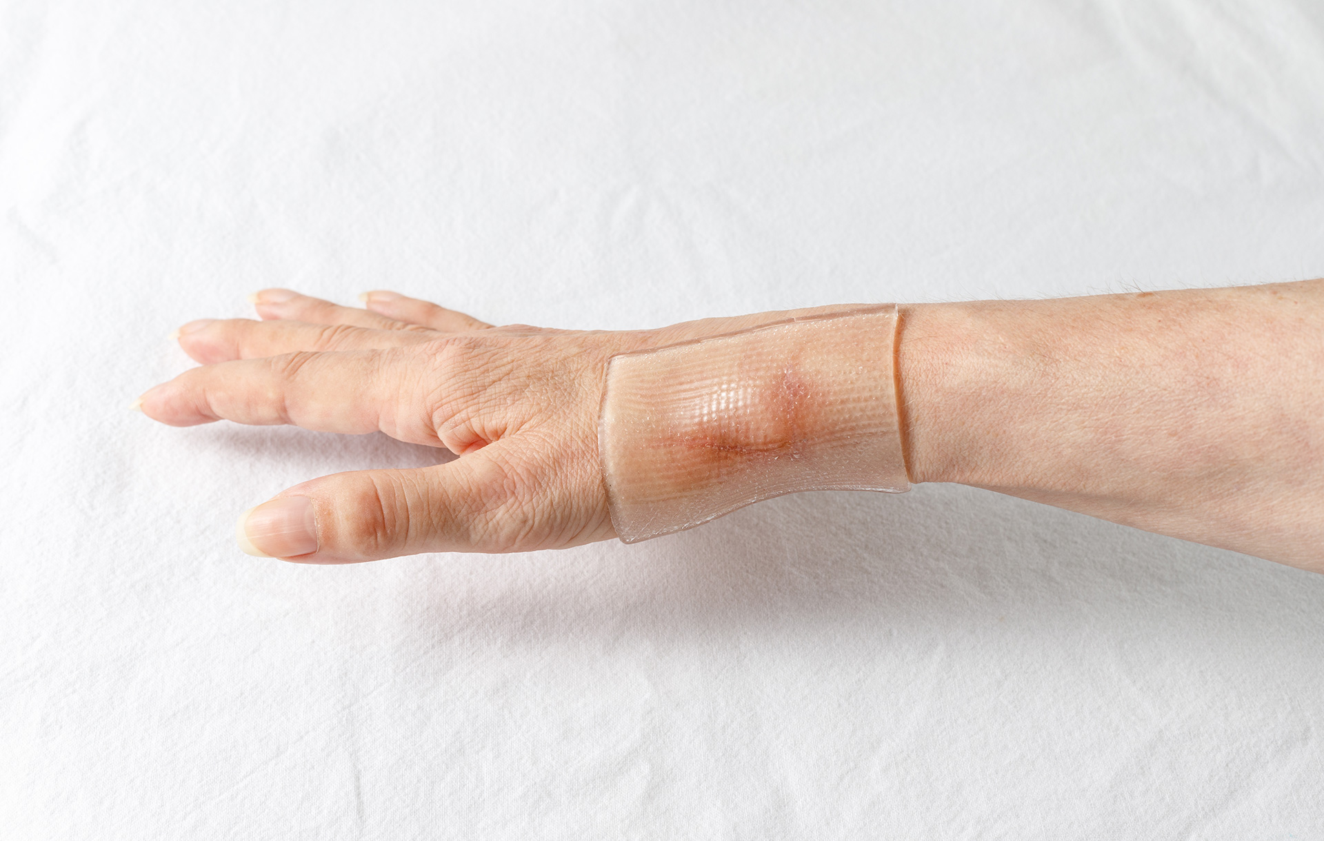
Skin injuries, infections, or diseases are a frequent occurrence within the general population. Oftentimes wound dressings or skin grafts are placed or implanted over the area of injury to assist with healing. The primary role of wound dressings is to isolate the site of injury from the environment and deliver drugs to allow the natural healing process to heal the wound. However, wound dressings require frequent exchanges irritating the injury site or pose an inherent risk to infection. Also, they do not play an active role in the healing process. Skin grafts on the other hand play an active role in the healing process. But many skin grafts either are limited in availability or pose a significant risk to rejection or infection.
To improve patient outcomes, ideally, wound dressings and skin grafts should be readily available, with low risk of rejection and infection and would not be require removal and replacement. Grafts or wound dressings composed of electrospun chitosan nanofibers, with innate antimicrobial and tissue regeneration properties have the potential to become the next generation wound healing scaffolds.
Skin Injuries and Diseases:
Common skin injuries or diseases include cuts, burns, and infections. Burn patients are frequently treated through autologous skin grafts.1 However, many autologous grafts have limitations in donor site availability. Other common skin grafts include xenograft of allografts; however, these types of grafts are prone to immune rejection and pose a risk of infection.2
Other common treatments of burns or wounds is through the application of a wound dressing that provides a moist environment and aims to restore tissue healing. The gold standard wound dressings include gauze, bandages, or cotton. These types of wound dressings simply isolate the wound, and often, lack antimicrobials, cause dehydration that delays healing, and requires frequent exchanges which could irritate the skin tissue.1 With these complications in mind, it is critical to design alternative wound dressings and skin grafts that prevent or reduce infection, reduce the need for reapplying dressings, control moisture balance, and improve tissue healing.
Chitosan:
A natural polymer of interest for many wound healing applications is chitosan. Chitosan is derived from its precursor, chitin, which is naturally found in crustacean species, or within the cell walls of fungi. The polymer is advantageous within wound healing because it has innate hemostatic, antimicrobial and anti-inflammatory properties.3 Furthermore, when applied to a wound, chitosan has been found to accelerate the wound healing process.4 As an added benefit, the chitosan polymer backbone can be crosslinked, which tunes the moisture retention of the polymer.
Electrospinning:
Electrospinning is a useful technology for developing polymer-based nanofibers that have a fiber diameter range between 100 nm to several microns. The electrospun nanofibers have many advantages for wound healing applications because the nanofibrous material mimics the structure of natural tissue that can promote cell adhesion, growth, and tissue production.5 Furthermore, electrospun fibers have a surface area to volume ratio that provides excellent gaseous exchange during healing as well as serving as a good source for drug delivery.6 The gaseous exchange rate can further be tunable by changing the fiber diameter, thereby controlling the pore size.
Developing Chitosan Electrospun Fiber Wound Dressings and Skin Grafts:
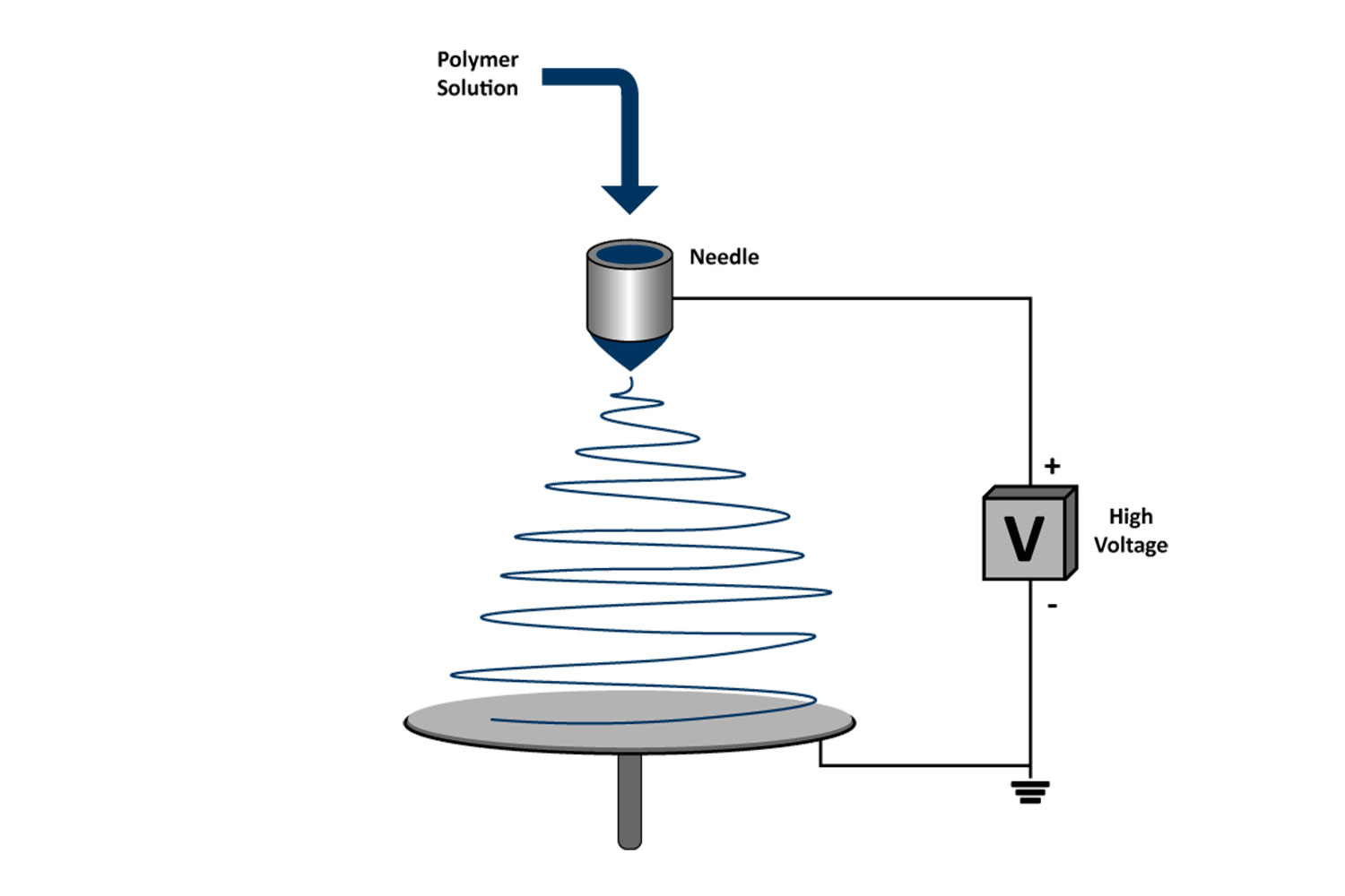
Chitosan is dissolved in green solvents to fabricate electrospun sheets composed of nanofibers. As the polymer solution is extruded through an emitter, the positively charged solution gets propelled towards a grounded or negatively charged target. During the time of flight, solvents evaporate, and the polymer is collected in the form of nanofibers (Figure 1).7 By depositing a significant amount of fibers on to the collector, sheets of chitosan nanofibers can be fabricated.
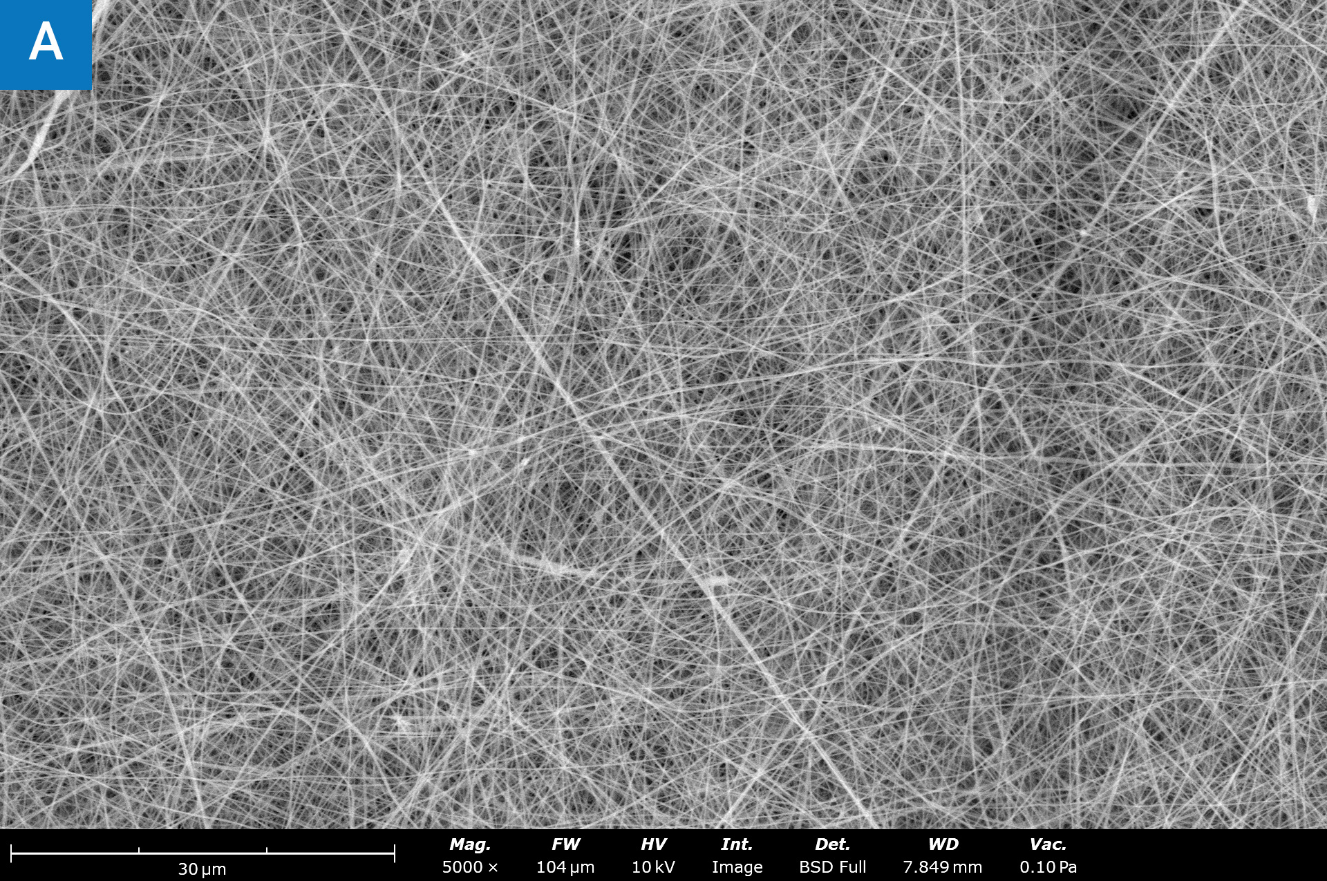
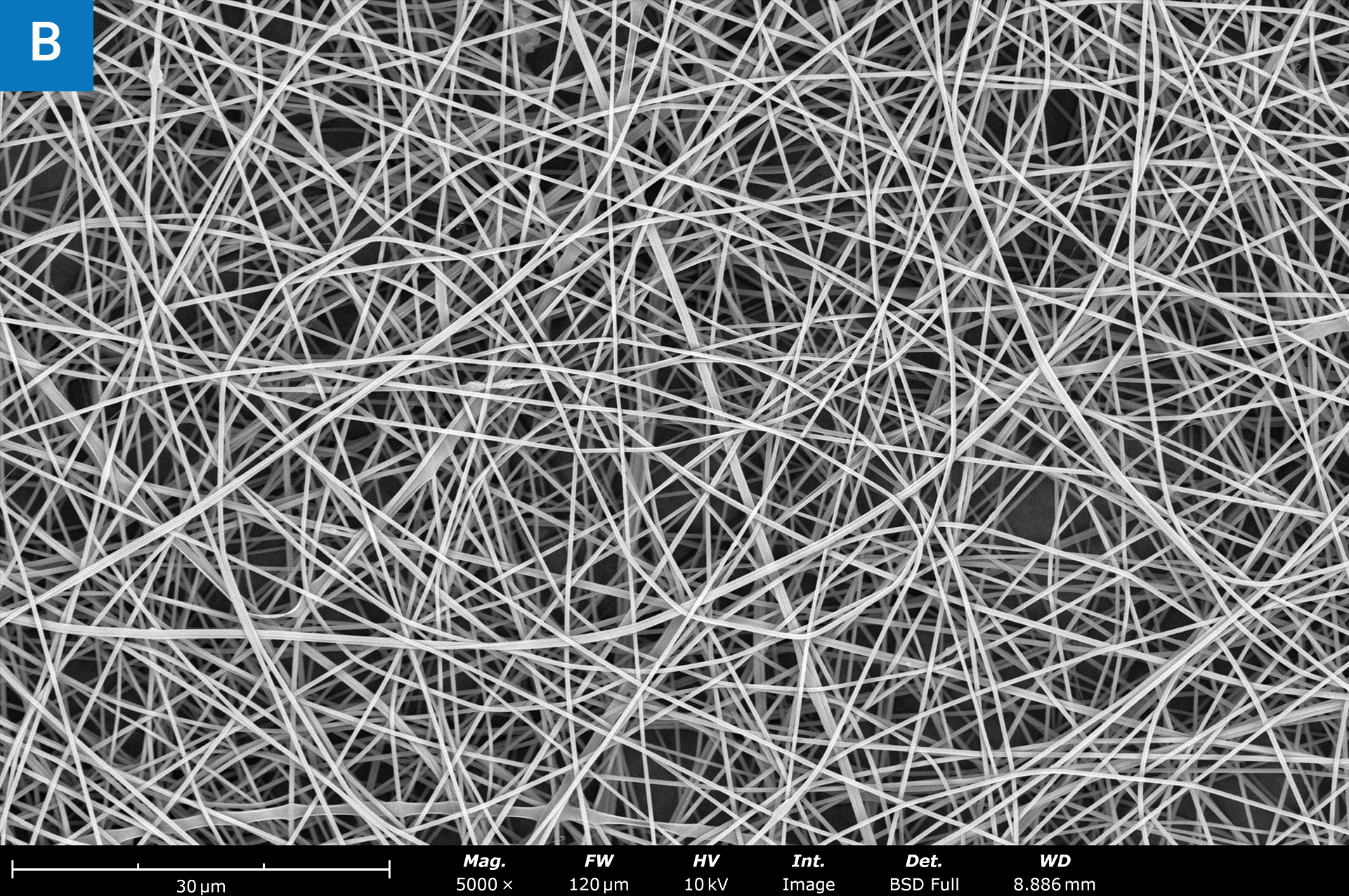
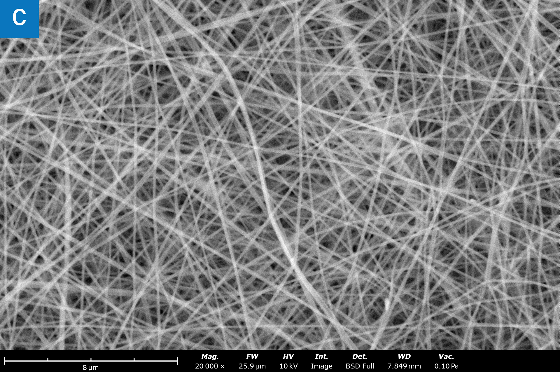
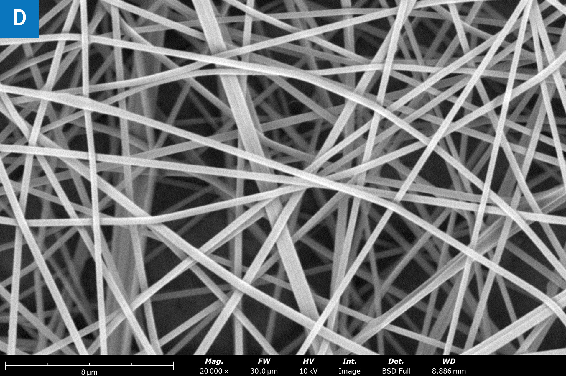
Scanning electron microscopy revealed the nanoscale structure of the smooth chitosan fibers (Figure 2). The average fiber diameter can be controlled by tuning the electrospinning formulation (Figure 2A, B). The pore sizes can be tuned by modulating the fiber diameter to optimize for drug delivery or gaseous exchange rates.
Conclusion
Designing next-generation wound dressings and skin grafts is critical for reducing infection, inflammation, retaining moisture, and inducing tissue healing and restoration. Electrospinning is an excellent technique that is used to make nanofibrous materials for wound healing applications. Developing chitosan electrospun fiber wound dressings mimics the structure of native tissue, has innate antimicrobial properties, can be crosslinked to tune water-retention, and serves as a proper scaffold for promoting proper skin healing.
References:
- Sheikholeslam, M., Wright, M. E., Cheng, N., Oh, H. H., Wang, Y., Datu, A. K., … & Jeschke, M. G. (2019). Electrospun polyurethane–gelatin composite: a new tissue-engineered scaffold for application in skin regeneration and repair of complex wounds. ACS biomaterials science & engineering, 6(1), 505-516. ↩︎
- Zhong, S. P., Zhang, Y. Z., & Lim, C. T. (2010). Tissue scaffolds for skin wound healing and dermal reconstruction. Wiley Interdisciplinary Reviews: Nanomedicine and Nanobiotechnology, 2(5), 510-525. ↩︎
- Bombin, A. D. J., Dunne, N. J., & McCarthy, H. O. (2020). Electrospinning of natural polymers for the production of nanofibres for wound healing applications. Materials Science and Engineering: C, 114, 110994. ↩︎
- Patrulea, V., Ostafe, V., Borchard, G., & Jordan, O. (2015). Chitosan as a starting material for wound healing applications. European Journal of Pharmaceutics and Biopharmaceutics, 97, 417-426. ↩︎
- Park, H., Patil, T. V., Dutta, S. D., Lee, J., Ganguly, K., Randhawa, A., … & Lim, K. T. (2024). Extracellular Matrix‐Bioinspired Anisotropic Topographical Cues of Electrospun Nanofibers: A Strategy of Wound Healing through Macrophage Polarization. Advanced Healthcare Materials, 2304114. ↩︎
- Jiang, X., Zeng, Y. E., Li, C., Wang, K., & Yu, D. G. (2024). Enhancing diabetic wound healing: Advances in electrospun scaffolds from pathogenesis to therapeutic applications. Frontiers in Bioengineering and Biotechnology, 12, 1354286. ↩︎
- Tindell, R. K., Busselle, L. P., & Holloway, J. L. (2023). Magnetic fields enable precise spatial control over electrospun fiber alignment for fabricating complex gradient materials. Journal of Biomedical Materials Research Part A, 111(6), 778-789. ↩︎